Table of Contents >> Show >> Hide
- Quick Map: The 3 Main Types of Retinal Detachment
- What the Retina Does (and Why Detachment Is a Big Deal)
- Type 1: Rhegmatogenous Retinal Detachment (RRD)
- Type 2: Tractional Retinal Detachment (TRD)
- Type 3: Exudative (Serous) Retinal Detachment
- Combined Detachments: When Life Refuses to Stay in One Category
- Symptoms: What People Usually Notice
- How Doctors Diagnose Retinal Detachment
- Treatments: From “Patch the Leak” to “Rebuild the Room”
- How Surgeons Choose the “Right” Treatment
- Recovery and Aftercare: What to Expect (General Guide)
- Outcomes, Complications, and Repeat Procedures
- Can You Prevent Retinal Detachment?
- When to Seek Emergency Care
- Real-World Experiences: What It Can Feel Like (and What People Often Wish They Knew)
- Conclusion
Retinal detachment is what happens when the retinathe light-sensing “film” lining the back of your eyelifts away from the tissue that supports and nourishes it. The retina is picky: once it loses contact, it can’t do its job well, and vision can drop fast. Think of it like wallpaper peeling off a wall. You don’t “just live with it” and hope it re-sticks by positive vibes.
Important: If you suddenly notice a shower of new floaters, flashes of light, or a shadow/“curtain” over part of your vision, treat it like a true eye emergency. (If you see a curtain, do not go shopping for drapesgo get evaluated.)
Quick Map: The 3 Main Types of Retinal Detachment
- Rhegmatogenous retinal detachment (RRD): A tear or hole lets fluid slip underneath the retina and lift it up. This is the most common type.
- Tractional retinal detachment (TRD): Scar tissue tugs the retina off the back wall of the eyeoften related to advanced diabetic eye disease.
- Exudative (serous) retinal detachment: Fluid builds up under the retina without a tear, usually from inflammation, abnormal blood vessels, or tumors.
Real life can be messier: some detachments are combined (for example, traction plus a tear), and treatment decisions depend on details like where the break is, whether the macula is involved, and what’s causing the traction or leakage.
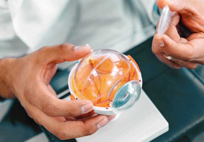
What the Retina Does (and Why Detachment Is a Big Deal)
Your retina converts light into signals your brain turns into vision. The center portion, the macula, handles sharp detailreading, driving, recognizing faces, and spotting that one typo your editor swears isn’t there.
When the retina detaches, it can become starved of oxygen and nutrients. The longer it stays detachedespecially if the macula is affectedthe higher the risk of lasting vision loss. That’s why doctors talk about urgency, not “wait-and-see.”
Type 1: Rhegmatogenous Retinal Detachment (RRD)
How it happens
“Rhegmatogenous” is a fancy way of saying: there’s a break in the retina (a tear or hole). Fluid from inside the eye can move through that break and collect under the retina, lifting it away from the underlying layer.
Common causes and risk factors
- Posterior vitreous detachment (PVD): As we age, the gel (vitreous) in the eye can pull away. Sometimes it tugs hard enough to tear the retina.
- Lattice degeneration: Thinning areas in the peripheral retina that are more prone to tearing.
- High myopia (nearsightedness): A longer eye shape can stretch and thin the retina.
- Eye trauma: Sports injuries, accidents, or blunt force can create tears.
- Prior eye surgery: Particularly after cataract surgery, the risk is higher than baseline.
- History of retinal tear/detachment in the other eye or family history.
A practical example
A 62-year-old notices quick flashes at night and new floaters after moving furniture. Two days later, they see a gray shadow creeping in from the side. In clinic, the doctor finds a retinal tear and fluid underneathclassic RRD storyline.
Type 2: Tractional Retinal Detachment (TRD)
How it happens
Instead of fluid sneaking under a tear, scar tissue forms on the retinal surface and contractslike shrink-wrap pulling tightlifting the retina off the back of the eye.
Common causes
- Proliferative diabetic retinopathy (PDR): Abnormal new blood vessels and fibrous tissue can form and pull.
- Other ischemic retinal diseases: Conditions that drive abnormal vessel growth and scarring.
- Prior inflammation or surgery that leads to membrane formation.
A practical example
A person with long-standing diabetes gradually notices distortion and blur rather than sudden flashes. Imaging shows traction bands pulling the retina, sometimes threatening or involving the macula. TRD can be quieter at first, but it is not “gentle.”
Type 3: Exudative (Serous) Retinal Detachment
How it happens
This type occurs when fluid leaks under the retina without a tear. The “leak” usually comes from inflammation, abnormal blood vessels, or a mass underneath the retina (such as a tumor). The retina is innocent hereit’s being flooded from below.
Common causes
- Inflammation (uveitis) or autoimmune conditions affecting the back of the eye.
- Abnormal blood vessel leakage (varies by condition).
- Tumors or other structural issues beneath the retina.
- Severe systemic conditions that change vascular leakage (rare, condition-specific).
A practical example
Someone develops blurry, wavy vision after significant inflammation in the eye. Exam and imaging show fluid under the retina but no tear. The treatment target becomes the underlying cause of leakagenot just “reattaching” the retina mechanically.
Combined Detachments: When Life Refuses to Stay in One Category
In advanced diabetic eye disease, traction can be present and a tear can form, creating a combined tractional-rhegmatogenous detachment. These often require more complex surgical planning because you’re dealing with both pulling forces and fluid through a break.
Symptoms: What People Usually Notice
Retinal detachment is often painlessannoyingly so, because pain would make it easier to take seriously. Common warning signs include:
- Sudden increase in floaters (specks, cobwebs, “pepper”)
- Flashes of light (especially in peripheral vision)
- A shadow/curtain moving across part of the visual field
- Sudden decrease in vision or distortion
Red flag combo: new flashes + lots of floaters + a creeping shadow. That’s a “call now / go now” situation.
How Doctors Diagnose Retinal Detachment
Diagnosis is usually made with a dilated eye exam. Your eye doctor uses bright lights and specialized lenses to look for tears, holes, and detached areas.
Additional tools may include:
- Ocular ultrasound if the view is blocked (for example, by bleeding in the vitreous).
- Optical coherence tomography (OCT) to assess the macula and fine retinal layers.
- Wide-field retinal imaging to document peripheral pathology.
These tests help answer the big clinical questions: Is there a break? Is the macula involved? Is there traction? What’s causing fluid?
Treatments: From “Patch the Leak” to “Rebuild the Room”
The goal is simple to say and hard to do: reattach the retina and prevent it from detaching again. The best approach depends on type, severity, and anatomy.
1) Treating retinal tears before detachment
If there’s a retinal tear but the retina isn’t detached (or only minimally), doctors may seal around the tear to stop fluid from slipping underneath.
- Laser photocoagulation: Tiny laser burns create a scar “weld” around the tear.
- Cryotherapy (cryopexy): A freezing treatment that also creates a sealing scar.
In other words: sometimes the best retinal detachment treatment is preventing the detachment from happening in the first place.
2) Pneumatic retinopexy (the “gas bubble + seal” approach)
This is often used for selected uncomplicated rhegmatogenous detachments. The surgeon injects a gas bubble into the eye; the bubble presses the retina back into place while laser or cryotherapy seals the tear.
Pros: Often done in-office; less invasive than some surgeries.
Trade-offs: Requires careful head positioning; not suitable for all tear locations or complex cases.
3) Scleral buckle (support from the outside)
A scleral buckle is a silicone band or sponge placed around the eye wall to gently indent it inward, reducing traction and helping the retinal break close. It’s commonly used in certain primary RRD cases and sometimes combined with other procedures.
Pros: Useful in specific anatomies; can preserve the natural lens in some situations.
Trade-offs: Surgical procedure; can change the eye’s shape slightly (which may affect glasses prescription).
4) Vitrectomy (cleaning out the pulling gel and membranes)
Pars plana vitrectomy removes the vitreous gel and any membranes tugging on the retina. The surgeon can flatten the retina, treat tears with laser/cryo, and then place a temporary internal support such as gas or silicone oil.
This is common for:
- More complex RRD
- Many tractional detachments
- Combined tractional-rhegmatogenous detachments
- Cases with significant scarring (like proliferative vitreoretinopathy)
5) Treating exudative detachment (fix the leak)
For exudative detachments, surgery to “patch a tear” may not helpbecause there isn’t one. Treatment focuses on the cause:
- Anti-inflammatory therapy when inflammation is driving leakage (treatment varies by diagnosis).
- Targeting abnormal vessels when leakage is vascular-driven (condition-specific).
- Tumor management when a mass is involved (handled by specialists).
The plan is often multidisciplinary: retina specialist, ocular oncology (if needed), and sometimes systemic evaluation.
How Surgeons Choose the “Right” Treatment
Retinal detachment surgery isn’t one-size-fits-all. Doctors decide based on a checklist of “retina reality” factors, such as:
- Type: rhegmatogenous vs tractional vs exudative (or combined)
- Macula status: macula-on vs macula-off (timing and urgency often differ)
- Location and number of tears (top vs bottom, single vs multiple)
- Lens status: natural lens vs prior cataract surgery
- Amount of scarring and traction (including proliferative vitreoretinopathy)
- Ability to posture (some approaches require strict positioning)
- Overall eye health (bleeding, inflammation, pressure issues)
That’s why two people with “retinal detachment” can have completely different repair plansand different recovery rules.
Recovery and Aftercare: What to Expect (General Guide)
Recovery depends on the procedure and what the surgeon placed inside the eye (gas, air, silicone oil, or nothing). Most people have multiple follow-ups early on, then spaced-out monitoring.
Vision recovery timeline
Vision often improves gradually over weeks to months. If the macula was detached, recovery can be slower and sometimes incomplete. It’s frustrating, but commonand it’s one reason early evaluation matters.
Positioning (yes, it matters)
If a gas bubble is used, you may be instructed to keep your head in a specific position to press the retina in the right place. This is temporary but important. It can feel like your neck is training for a very niche Olympics event.
Altitude rules (seriously, don’t improvise)
If you have a gas bubble, you may need to avoid flying and high altitudes until the bubble is gone. Altitude changes can expand gas and raise eye pressure. Your surgeon will tell you what’s safe and when.
Common short-term experiences
- Scratchy sensation, watering, or mild discomfort
- Temporary blurriness (especially with gas)
- Seeing a shifting line or circle (the bubble edge) as it slowly absorbs
- Activity restrictions for a period (your surgeon sets the rules)
Pro tip: Follow your post-op instructions exactly. Retinas do not respond well to “I made my own plan.”
Outcomes, Complications, and Repeat Procedures
Modern retinal detachment repair often succeeds at reattaching the retina, but outcomes vary based on timing, type, and complexity. Some people need more than one procedure, especially in complex detachments or when scar tissue develops.
Possible complications can include cataract progression (especially after vitrectomy), elevated eye pressure, infection (rare but serious), bleeding, or recurrent detachment. Your team watches for these closely in follow-up visits.
Can You Prevent Retinal Detachment?
You can’t fully “life-hack” your way out of retinal detachment risk, but you can reduce the odds of a bad outcome:
- Get urgent evaluation for new flashes, floaters, or a curtain/shadow.
- Manage diabetes well and keep up with diabetic eye screening if you have diabetes.
- Protect your eyes during sports or high-risk work.
- Keep routine eye exams if you’re highly nearsighted or have a history of retinal issues.
- Know your risk if you’ve had cataract surgery or a detachment in the other eye.
When to Seek Emergency Care
Call an eye doctor urgently or go to emergency care if you have:
- Sudden flashes of light
- A sudden “storm” of new floaters
- A shadow or curtain over any part of your vision
- Sudden, unexplained vision loss
Better to be told “good news, it’s not a detachment” than to wait and lose treatable vision.
Real-World Experiences: What It Can Feel Like (and What People Often Wish They Knew)
This section is based on common patient-reported experiences and typical clinical pathways. Everyone’s eyesand emotionsare different.
1) The symptoms can feel weirdly harmless at first. Many people describe the early stage as “annoying,” not “terrifying.” A few floaters can look like dust on a camera lens. Flashes can be mistaken for a phone screen lighting up, a car headlight, or that one ceiling bulb that always flickers. The problem is that your brain is excellent at normalizing strange visual stuffuntil a shadow starts creeping in and you realize your eye is not auditioning for a special-effects movie.
2) The emotional whiplash is real. People often go from “I’m probably overreacting” to “Why is everyone saying the word emergency?” in under an hour. There’s a particular kind of stress that comes with a painless crisis: you feel okay, but the situation isn’t okay. It’s common to feel guilty for waiting, even if you only waited overnight. If you’re reading this for yourself or someone you care about, take the guilt off the table. Use that energy to get evaluated and follow the plan.
3) The exam can be intense but not mysterious. A dilated retinal exam involves bright lights, lots of looking around, and instructions like “look up… now left… now down.” It may feel long, but it’s purposeful: the doctor is hunting for the tear, mapping the detachment, and deciding how urgent and complex the repair will be. Some people remember thinking, “I didn’t know my eye had this many directions.” (It does. It’s basically a tiny globe with opinions.)
4) Treatment day often feels faster than the build-up. If the plan is a laser procedure or cryotherapy for a tear, people frequently report surprise at how quick it is. For surgery (pneumatic retinopexy, scleral buckle, vitrectomy, or a combination), the day can be a blurarrive, prep, procedure, recovery, instructions. Many patients say the hardest part is not the procedure itself, but the mental load: remembering the rules, managing follow-ups, and adjusting expectations about vision.
5) The “bubble life” can be the strangest part. If a gas bubble is used, people often describe seeing a moving line like a tiny horizon inside the eye. As the bubble shrinks, that line drops lower, sometimes creating a wobbly “fishbowl” effect. Reading can be awkward, depth perception can be off, and patience becomes a daily assignment. Positioning (face-down or side positioning) can be annoying, but many people find it easier once they set up a comfortable stationpillows, audiobooks, podcasts, and a reminder that this is temporary and purposeful.
6) Recovery is rarely a straight lineand that’s normal. Some days vision feels clearer; other days it feels foggy. Dryness, mild irritation, and light sensitivity are common early on. People often worry that every odd sensation means failure. Most of the time, it’s just healing doing healing things. The key experience many patients share: follow-up visits are reassuring because they turn vague worries into measurable factsretina attached, pressure okay, inflammation improving, next steps clear.
7) People often wish they’d known how common “help” is. Needing rides, taking time off work, asking someone to read small print, or simply having a friend sit with youthese are normal parts of the process. Retinal detachment can feel isolating because it’s happening in one eye, but it affects your whole routine. The most helpful mindset shift patients describe is treating recovery like a short-term project with a schedule: meds, positioning (if required), rest, follow-up, repeat. Not glamorous, but very doable.
Bottom line: Retinal detachment is serious, but it’s also treatableespecially when caught early. If you notice warning signs, getting evaluated quickly is the most powerful move you can make.
Conclusion
Retinal detachment comes in three main typesrhegmatogenous, tractional, and exudativeand each has a different “why,” which shapes the “how” of treatment. Rhegmatogenous detachments usually start with a tear; tractional detachments are pulled by scar tissue (often linked to diabetes); and exudative detachments result from leakage under the retina without a break. Today’s treatmentsfrom laser and cryotherapy to pneumatic retinopexy, scleral buckle, and vitrectomyare highly specialized, and outcomes are best when care happens quickly.
If you remember only one thing: sudden floaters, flashes, or a curtain over vision should never be ignored. In the retina world, speed is not panicit’s strategy.