Table of Contents >> Show >> Hide
- What Is Plantar Vein Thrombosis?
- Why Plantar Vein Thrombosis Is Often Misdiagnosed
- Common Symptoms of Plantar Vein Thrombosis
- What Causes Plantar Vein Thrombosis?
- How Plantar Vein Thrombosis Is Diagnosed
- Differential Diagnosis: Conditions That Can Look Similar
- Treatment for Plantar Vein Thrombosis
- Recovery: What to Expect
- Prevention: Lowering the Risk of Future Clots
- Living With Plantar Vein Thrombosis: Practical Experience Notes
- Conclusion
Plantar vein thrombosis is not the foot problem most people expect when heel or arch pain suddenly shows up. Most of us blame the usual suspects: new shoes, too much walking, a heroic but regrettable gym session, or the classic “I stepped weird and now my foot has opinions.” But in rare cases, pain in the sole of the foot can come from a blood clot inside one of the plantar veins.
That is plantar vein thrombosis, often shortened to PVT. It is uncommon, underrecognized, and easy to mistake for plantar fasciitis, tendon irritation, a stress fracture, or general foot inflammation. The tricky part is that symptoms can be annoyingly ordinary: pain, swelling, tenderness, and difficulty walking. The important part is that a clot deserves medical attention because venous clots can sometimes extend, recur, or become part of a more serious venous thromboembolism picture.
This guide explains the symptoms, diagnosis, treatment options, risk factors, and real-world patient experience around plantar vein thrombosis in plain American English. No medical-school decoder ring required.
Medical note: This article is for educational purposes only. It is not a diagnosis, treatment plan, or replacement for care from a licensed healthcare professional. If you have sudden foot swelling, unexplained severe foot pain, chest pain, shortness of breath, fainting, or coughing blood, seek urgent medical care.
What Is Plantar Vein Thrombosis?
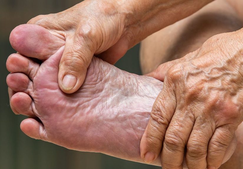
Plantar vein thrombosis is a blood clot that forms in one or more veins on the bottom of the foot. The plantar veins help move blood from the foot back toward the heart. When a clot blocks or partly blocks one of these veins, blood flow becomes disrupted, local tissues may become irritated, and pain can develop in the heel, arch, or sole.
The word “plantar” refers to the sole of the foot. The word “thrombosis” means clot formation inside a blood vessel. Put them together, and plantar vein thrombosis means “a clot in a vein under the foot.” Not glamorous, but definitely important.
PVT is considered rare, but many experts believe it may be missed because standard lower-leg ultrasound exams do not always include the veins in the sole. In other words, the clot may be hiding in the basement while everyone is checking the upstairs hallway.
Why Plantar Vein Thrombosis Is Often Misdiagnosed
Most people with foot pain do not have plantar vein thrombosis. Common conditions such as plantar fasciitis, Achilles or posterior tibial tendon problems, metatarsalgia, Morton’s neuroma, arthritis, and stress fractures are far more common. That is one reason PVT can be overlooked.
Another reason is that PVT often causes nonspecific symptoms. A patient may say, “My arch hurts,” “My heel feels swollen,” or “It hurts when I stand.” Those complaints sound very similar to ordinary overuse injuries. Unless the clinician suspects a vascular cause, the plantar veins may not be examined closely.
PVT vs. Plantar Fasciitis
Plantar fasciitis usually causes sharp heel pain, often worse with the first steps in the morning or after sitting. PVT can also cause heel or arch pain, but it may come with swelling, warmth, a heavy feeling in the foot, or pain that feels deeper and more localized along the medial or lateral arch.
A helpful clue is persistence despite typical treatment. If stretching, shoe changes, rest, and anti-inflammatory measures do not help, or if swelling and vascular risk factors are present, clinicians may consider imaging beyond the usual musculoskeletal workup.
Common Symptoms of Plantar Vein Thrombosis
The symptoms of plantar vein thrombosis can vary, but the most reported signs include:
- Pain in the sole of the foot
- Heel pain or arch pain
- Pain that worsens when standing or walking
- Localized swelling in the foot
- A heavy or full feeling in the affected foot
- Tenderness along the plantar arch
- Warmth, redness, or skin discoloration in some cases
- Difficulty bearing weight
- Symptoms affecting one foot more than the other
Many cases appear on one side only. A person may feel fine in the morning, then notice that one foot becomes painful after activity. Others describe a deep ache that does not match the usual “pulled muscle” feeling.
When Symptoms Require Urgent Attention
Seek prompt medical care if foot pain or swelling is unexplained, sudden, one-sided, worsening, or associated with known clotting risk factors. Seek emergency help immediately if you develop symptoms that may suggest pulmonary embolism, including sudden shortness of breath, chest pain that worsens with deep breathing, rapid heartbeat, fainting, dizziness, or coughing up blood.
What Causes Plantar Vein Thrombosis?
Like other venous clots, plantar vein thrombosis may develop when one or more parts of “Virchow’s triad” are present: slow blood flow, vessel wall injury, and increased tendency of the blood to clot. That sounds fancy, but it simply means clots are more likely when blood sits still, a vein is irritated or injured, or the blood is unusually clot-prone.
Possible triggers and risk factors include:
- Recent foot or ankle injury
- Foot surgery or lower-limb surgery
- Prolonged immobilization, such as bed rest or a walking boot
- Long-distance travel with extended sitting
- Cancer or cancer treatment
- Pregnancy or the postpartum period
- Estrogen-containing birth control or hormone therapy
- Inherited clotting disorders
- Previous deep vein thrombosis or pulmonary embolism
- Obesity
- Smoking
- Inflammatory or chronic medical conditions
- Mechanical strain from intense walking, running, or prolonged standing
Sometimes no obvious cause is found. That can be frustrating, but it does happen with venous clots. A healthcare provider may call this “unprovoked” thrombosis and may consider additional evaluation depending on the patient’s age, medical history, recurrence risk, and family history.
How Plantar Vein Thrombosis Is Diagnosed
Diagnosis usually begins with a medical history and physical exam. The clinician will ask when the pain started, where it hurts, whether swelling is present, whether symptoms worsen with standing, and whether you have risk factors for blood clots.
Because PVT can mimic common foot conditions, imaging is often necessary. A careful diagnosis matters because treatment for a clot is different from treatment for ordinary plantar fasciitis. Stretching your calf is helpful for many heel pain problems, but it will not magically negotiate with a blood clot.
Duplex Ultrasound
Duplex ultrasound is commonly used to evaluate venous thrombosis. It uses sound waves to look at blood flow and vein compressibility. In a clot-free vein, the vessel usually compresses under gentle pressure from the ultrasound probe. A thrombosed vein may not compress normally and may show clot material inside the vessel.
For plantar vein thrombosis, the key is that the ultrasound exam must include the plantar veins. Standard DVT ultrasound protocols often focus on the calf, thigh, or pelvis, not the bottom of the foot. If PVT is suspected, the ordering clinician may need to specifically request evaluation of the medial and lateral plantar veins.
MRI
Magnetic resonance imaging, or MRI, can be very helpful when ultrasound is inconclusive or when the diagnosis is not suspected at first. MRI may show an enlarged vein, clot signal within the vessel, surrounding soft-tissue swelling, and other features that distinguish PVT from tendon injury, neuroma, bursitis, or stress fracture.
MRI is also useful because foot pain has many possible causes. One scan may help evaluate bones, joints, tendons, fascia, nerves, and soft tissues. That makes MRI a valuable “detective tool” when symptoms are stubborn or unusual.
D-Dimer Blood Test
A D-dimer test may be used in some suspected clotting situations. D-dimer is a blood marker that can rise when clots form and break down. A normal D-dimer may help rule out clotting in low-risk patients, but it is not specific. Many things can raise D-dimer, including infection, inflammation, surgery, trauma, pregnancy, and cancer. For plantar vein thrombosis, imaging remains central.
Differential Diagnosis: Conditions That Can Look Similar
Because plantar vein thrombosis is uncommon, clinicians usually consider more common diagnoses first. Similar conditions include:
- Plantar fasciitis: inflammation or degeneration of the plantar fascia, often causing first-step heel pain.
- Stress fracture: tiny bone injury from repetitive load, often worse with activity.
- Tendinopathy: tendon irritation, especially around the ankle or arch.
- Morton’s neuroma: nerve irritation commonly causing forefoot pain, burning, or numbness.
- Metatarsalgia: pain under the ball of the foot.
- Crystal arthritis: gout or pseudogout causing sudden pain and inflammation.
- Infection: redness, warmth, swelling, fever, or skin breaks may point toward infection.
- Nerve entrapment: such as Baxter’s nerve irritation, which may mimic heel pain.
The overlap is exactly why persistent, one-sided, swollen, or unusual foot pain should be evaluated rather than self-diagnosed from a search engine at 1:00 a.m. Search engines are great at panic; doctors are better at context.
Treatment for Plantar Vein Thrombosis
Treatment depends on the size and location of the clot, symptoms, risk factors, risk of clot extension, bleeding risk, and whether other veins are involved. There is no single universal treatment plan for every PVT case because the condition is rare and formal guidelines are limited. However, clinicians often borrow principles from lower-extremity DVT management.
Anticoagulant Medication
Anticoagulants are medications that reduce the blood’s ability to clot. People often call them “blood thinners,” although they do not actually make blood watery. They help prevent the existing clot from growing and reduce the risk of new clots while the body gradually breaks down the clot.
Common options may include direct oral anticoagulants, low-molecular-weight heparin, unfractionated heparin, fondaparinux, or warfarin. The right choice depends on kidney function, liver function, pregnancy status, cancer history, medication interactions, cost, bleeding risk, and clinician judgment.
Some patients may need treatment for several weeks to several months. Others may need longer treatment if the clot is recurrent, unprovoked, associated with cancer, or linked to a strong thrombophilia. This decision should be individualized.
Pain Control and Supportive Care
Supportive care may include rest, activity modification, elevation, and pain control. Some clinicians may recommend acetaminophen for pain. Nonsteroidal anti-inflammatory drugs, such as ibuprofen or naproxen, may not be appropriate for everyone, especially people taking anticoagulants, because they can increase bleeding risk. Always ask your healthcare provider before combining medications.
Footwear support may help reduce mechanical stress while symptoms improve. In some cases, temporary reduction of high-impact activity is reasonable. That does not mean becoming a couch statue forever. It means letting the foot calm down while the treatment plan does its job.
Compression and Follow-Up Imaging
Depending on the case, clinicians may recommend compression therapy or follow-up duplex ultrasound to check whether the clot is stable, improving, or extending. Follow-up is especially important if symptoms worsen or if there is concern about clot propagation into larger veins.
When Procedures Are Considered
Procedures such as thrombolysis or thrombectomy are generally reserved for selected high-risk clotting situations, such as extensive limb-threatening thrombosis or serious pulmonary embolism. Isolated plantar vein thrombosis is usually managed medically, but a vascular specialist may become involved when the case is complicated, recurrent, or associated with other venous disease.
Recovery: What to Expect
Recovery varies. Some people notice pain improvement within days or weeks after treatment begins. Others have lingering soreness, swelling, or activity limitation for longer. Imaging may show partial or complete reopening of the vein over time, but symptom recovery and imaging recovery do not always move at the exact same speed.
During recovery, patients should follow medication instructions carefully. Missing anticoagulant doses can reduce protection. Taking extra doses can increase bleeding risk. This is not the time for freestyle pharmacology.
Patients taking anticoagulants should ask their healthcare provider about signs of bleeding, medication interactions, dental procedures, sports participation, and what to do if they fall or injure themselves.
Prevention: Lowering the Risk of Future Clots
Not every case of plantar vein thrombosis can be prevented, but several habits may reduce clot risk:
- Move regularly during long travel or desk work.
- Stand, walk, or do ankle pumps every one to two hours during long trips.
- Follow post-surgical movement and medication instructions.
- Discuss clot risk before surgery, pregnancy, or hormone therapy.
- Maintain a healthy weight when possible.
- Avoid smoking.
- Stay hydrated during travel and illness.
- Wear prescribed compression garments if recommended.
- Report new one-sided swelling or unexplained pain promptly.
If you have a personal or family history of clots, your doctor may recommend a more specific prevention plan. That plan may change during high-risk periods such as hospitalization, surgery, long-distance travel, cancer treatment, pregnancy, or postpartum recovery.
Living With Plantar Vein Thrombosis: Practical Experience Notes
People dealing with plantar vein thrombosis often describe the experience as confusing because the pain feels like a “regular foot problem” at first. A person may buy new insoles, roll the foot on a frozen water bottle, stretch the calf, change shoes, and still feel that something is not right. The turning point is often the combination of one-sided pain, swelling, heaviness, and symptoms that do not behave like ordinary plantar fasciitis.
One practical lesson is to track symptoms clearly. Instead of saying only “my foot hurts,” write down where it hurts, when it started, whether swelling is present, whether the foot feels warm, whether walking makes it worse, and whether you recently traveled, had surgery, wore a boot, injured the foot, changed hormones, or spent long hours sitting. These details help clinicians decide whether imaging should include the plantar veins.
Another common experience is frustration after a normal first evaluation. A person may have an X-ray that shows no fracture or an exam that points toward plantar fasciitis. That does not mean the pain is imaginary. It may mean the right structure has not been examined yet. If symptoms persist or worsen, follow-up matters. Medicine is sometimes less like flipping a light switch and more like detective work with better shoes.
Patients on anticoagulants often learn that treatment is not just “take a pill and forget it.” They need to understand bleeding precautions, drug interactions, missed-dose instructions, and when to call the doctor. Even simple choices, such as taking ibuprofen for foot pain, may need medical approval. People who enjoy contact sports, heavy lifting, trail running, or adventurous hobbies should ask about safe activity while taking anticoagulants.
Footwear also becomes surprisingly important. During recovery, many patients do better with supportive shoes, cushioned soles, and reduced high-impact activity. Barefoot walking on hard floors may irritate symptoms. Long standing may trigger aching. A gradual return to walking, work shifts, or exercise is usually more sensible than declaring war on the treadmill on day three.
The emotional side is real too. Hearing “blood clot” can be scary, especially when online searches jump straight to worst-case scenarios. A balanced view helps: plantar vein thrombosis is uncommon and treatable, but it should be respected. The goal is not panic; the goal is timely diagnosis, appropriate treatment, and smart follow-up.
For many people, the biggest takeaway is self-advocacy. If your foot pain is severe, one-sided, swollen, unusual, or connected to clotting risk factors, say so clearly. Ask whether a vascular cause should be considered. Ask whether imaging included the plantar veins. Ask what symptoms should send you to urgent care. Good questions do not annoy good clinicians; they sharpen the picture.
Conclusion
Plantar vein thrombosis is a rare but important cause of heel, arch, or sole pain. Because it can look like plantar fasciitis or other common foot problems, it may be missed unless clinicians consider a vascular diagnosis. The most useful clues include one-sided plantar foot pain, swelling, heaviness, pain with standing or walking, and clotting risk factors such as recent surgery, injury, immobility, long travel, pregnancy, hormone therapy, cancer, or a history of blood clots.
Diagnosis usually depends on imaging, especially duplex ultrasound that specifically evaluates the plantar veins or MRI when the diagnosis is uncertain. Treatment may include anticoagulant medication, pain control, activity modification, compression when appropriate, and follow-up imaging. The best plan is individualized because PVT is uncommon and treatment decisions must balance clot risk with bleeding risk.
If your foot pain is acting suspiciously dramatic, do not ignore it. Most heel pain is not a blood clot, but unexplained swelling, severe one-sided pain, warmth, redness, or symptoms linked to clot risk deserve medical attention. Your feet carry you everywhere. When one of them sends a serious memo, read it.