Table of Contents >> Show >> Hide
- Quick refresher: what PID actually is
- So… does PID show up on ultrasound?
- What ultrasound can show in PID
- What ultrasound usually can’t show (and why PID can be missed)
- Why clinicians order an ultrasound when PID is suspected
- What other tests are used along with (or instead of) ultrasound?
- If your ultrasound is “normal,” could you still have PID?
- What treatment looks like (briefly, because this isn’t a prescription pad)
- When to seek urgent care
- FAQ: fast answers to common ultrasound-and-PID questions
- Bottom line
- Real-World Experiences: What People Notice (and What Clinicians See)
- Experience #1: “My ultrasound was normal, but my doctor still said PID.”
- Experience #2: “The ultrasound tech spent forever on one side… and everyone got quiet.”
- Experience #3: “I felt embarrassedlike this is my fault.”
- Experience #4: “The antibiotics helped… but I’m still anxious.”
- Experience #5: “I didn’t realize pelvic pain could be so complicated.”
If you’re hoping an ultrasound will give you a crisp, dramatic “YES, THIS IS PID” stamplike a movie detective
slamming a folder on the tableI have gently disappointing news: pelvic inflammatory disease (PID) is often a clinical diagnosis,
and ultrasound is more like a helpful sidekick than the main character.
That said, ultrasound can absolutely show cluesespecially when PID has progressed or complications are present.
The real trick is understanding what ultrasound can and can’t see, and why a “normal” scan doesn’t automatically mean “nothing’s wrong.”
Quick refresher: what PID actually is
PID is an infection/inflammation of the upper reproductive tracttypically involving the uterus, fallopian tubes, and/or ovaries.
It’s commonly linked to sexually transmitted infections (STIs) like chlamydia and gonorrhea, though other bacteria can be involved too.
Left untreated, PID can lead to scarring and serious long-term problems like infertility, ectopic pregnancy, chronic pelvic pain, and abscesses.
So… does PID show up on ultrasound?
Sometimes. Ultrasound may show findings that support PIDespecially when inflammation is significant or when complications like a
tubo-ovarian abscess (TOA) develop. But PID can also be present with no obvious ultrasound findings, particularly in early or mild cases.
The headline you came for
- Ultrasound can support a PID diagnosis when it shows classic inflammatory changes (especially in the fallopian tubes).
- Ultrasound cannot reliably rule out PIDa normal scan doesn’t exclude it.
- Ultrasound is most useful for spotting complications (like TOA) or ruling out look-alikes (like ectopic pregnancy or ovarian torsion).
What ultrasound can show in PID
Ultrasound findings in PID tend to be more visible when the fallopian tubes and ovaries are clearly affected or when pus/fluid collections form.
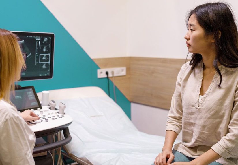
Transvaginal ultrasound (probe in the vagina) generally provides better detail than transabdominal ultrasound (probe on the belly).
Common PID ultrasound findings clinicians look for
- Thickened, fluid-filled fallopian tubes (a big supportive clue, sometimes described as “dilated tubes” or “pyosalpinx” if infected fluid is present)
- Free pelvic fluid (fluid in the pelvis, sometimes in the cul-de-sac/pouch behind the uterus)
- Tubo-ovarian complex or tubo-ovarian abscess (TOA) (a mass-like inflammatory collection involving tube/ovary)
- Increased blood flow (hyperemia) on Doppler suggesting active inflammation/infection
- Characteristic signs described in ultrasound literature (for example, a “cogwheel” appearance in cross-section of an inflamed tube)
Some clinical guidelines explicitly list imaging findingslike thickened, fluid-filled tubes (with or without free fluid or TOA) and Doppler evidence of tubal hyperemiaas
supportive criteria for PID when the diagnosis is uncertain. In other words: ultrasound can add confidence, but it’s not the whole story.
What ultrasound usually can’t show (and why PID can be missed)
Ultrasound is great at showing structures and fluid collections. PID, however, can start as inflammation and infection on a microscopic level
well before the tubes look enlarged or fluid-filled. That’s why you’ll see a recurring theme in reputable medical references:
absence of ultrasound findings does not exclude PID.
Reasons PID may not show on ultrasound
- Early infection hasn’t produced visible tubal swelling or fluid yet.
- Mild PID may cause pain and tenderness without dramatic structural changes.
- Ultrasound is operator-dependent: image quality, technique, and experience matterespecially for subtle tubal findings.
- Symptoms overlap with many other causes of pelvic pain, so ultrasound is often used to triage and rule out emergencies rather than “confirm PID.”
Why clinicians order an ultrasound when PID is suspected
In real clinics and ERs, ultrasound is often ordered for three practical reasons:
1) To look for complications (especially tubo-ovarian abscess)
A tubo-ovarian abscess can be serious and may change management (hospital admission, IV antibiotics, and sometimes drainage or surgery).
Ultrasound is commonly used to identify TOA and to help guide next steps.
2) To rule out conditions that can look like PID but need different urgent treatment
Pelvic pain is an overachiever. It can be caused by many things, including:
- Ectopic pregnancy (a pregnancy test is critical in evaluation)
- Ovarian torsion
- Ruptured ovarian cyst
- Appendicitis or other abdominal pathology
Ultrasound helps clinicians rapidly evaluate the uterus and ovaries and decide whether PID is most likelyor whether another diagnosis needs urgent attention.
3) To gather supportive evidence when the diagnosis is uncertain
PID is frequently diagnosed based on symptoms and pelvic exam findings (like cervical motion tenderness, uterine tenderness, or adnexal tenderness),
because delaying treatment can increase the risk of complications. Imaging and lab tests can support the diagnosis, but clinicians often treat based on clinical suspicion rather than “wait for perfect proof.”
What other tests are used along with (or instead of) ultrasound?
Since PID doesn’t have a single “magic test,” clinicians typically combine several pieces of information:
- History and symptoms (pelvic/lower abdominal pain, abnormal discharge, bleeding, fever, pain with sex, etc.)
- Pelvic exam (cervical motion tenderness, uterine or adnexal tenderness)
- Pregnancy test (to rule out ectopic pregnancy)
- STI testing (chlamydia/gonorrhea, and often additional screening)
- Blood tests (sometimes markers of inflammation/infection)
-
More advanced evaluation in selected cases:
MRI, endometrial biopsy, or laparoscopy (rarely, because it’s invasive)
If your ultrasound is “normal,” could you still have PID?
Yes. This is one of the most confusing parts for patients:
you can have PID with a normal ultrasound.
The diagnosis may still be made based on symptoms, exam findings, risk factors, and lab results.
Think of ultrasound like a flashlight in a foggy room. It can illuminate big stuff (like an abscess), but it might not reveal early inflammation.
That’s why major clinical references emphasize treating suspected PID promptly rather than waiting for imaging confirmation.
What treatment looks like (briefly, because this isn’t a prescription pad)
PID is typically treated with antibiotics. Treatment is often started right away when PID is suspected, even before all test results return.
Severe cases, pregnancy, inability to tolerate oral meds, or concern for abscess can lead to hospital treatment with IV antibiotics.
Sexual partners may need evaluation/treatment to prevent reinfection.
When to seek urgent care
Pelvic pain can be serious. Seek urgent evaluation if you have:
- Severe or worsening pelvic/lower abdominal pain
- Fainting, dizziness, shoulder pain (possible ectopic pregnancy concern)
- High fever, vomiting, or signs of severe illness
- Known pregnancy with pelvic pain
- Inability to keep fluids/medications down
FAQ: fast answers to common ultrasound-and-PID questions
Is transvaginal ultrasound better for PID?
Generally, yes. Transvaginal ultrasound usually provides clearer images of pelvic organs and can better detect subtle tubal or adnexal abnormalities.
Can ultrasound detect PID scarring?
Sometimes it may suggest chronic changes (like hydrosalpinxfluid-distended tube), but ultrasound isn’t a perfect “scar detector.”
Scarring and adhesions can be difficult to visualize directly. The bigger concern is that scarring can exist even after symptoms improve.
Can an ultrasound show endometritis (uterine lining inflammation)?
Ultrasound may show nonspecific findings (like fluid) but endometritis is often diagnosed based on clinical evaluation and, in selected cases, biopsy
not ultrasound alone.
Bottom line
PID can show on ultrasoundbut it doesn’t always. Ultrasound is most helpful for finding complications like tubo-ovarian abscess
and for excluding other urgent causes of pelvic pain. If symptoms and exam findings suggest PID, clinicians often treat promptly because waiting for a “perfect” scan can be risky.
Real-World Experiences: What People Notice (and What Clinicians See)
Let’s talk about the part medical websites can’t fully capture: what it actually feels like to go through the “Do I have PID?” workupespecially when an ultrasound is involved.
Spoiler: it can be frustratingly indirect. PID doesn’t always announce itself with a neon sign, and patients often come away thinking,
“Wait… if the ultrasound was normal, why am I still being treated?”
Experience #1: “My ultrasound was normal, but my doctor still said PID.”
This happens a lot. People show up with pelvic pain, maybe abnormal discharge, maybe pain during sex, maybe spotting that’s new for them.
They get a transvaginal ultrasound, and the report says something like “no acute abnormality” or “unremarkable.” Then the clinician says,
“I still think we should treat for PID.” That can feel like medicine is guessing.
But here’s the logic patients often don’t hear out loud: early PID can cause significant pain and tenderness before the fallopian tubes look swollen or fluid-filled.
The clinician is using the pelvic exam findings, STI risk profile, and symptom pattern to make a call. Ultrasound, in this scenario, is doing a different job
mainly ruling out emergencies (like torsion) and looking for obvious complications.
The “normal ultrasound” isn’t a dismissal; it’s one piece of the puzzle that simply didn’t light up.
Experience #2: “The ultrasound tech spent forever on one side… and everyone got quiet.”
When ultrasound does show something, it can shift the mood quicklyespecially if there’s a suspected tubo-ovarian abscess.
Patients often describe the exam as suddenly more thorough, with repeated angles and Doppler checks. Clinicians may move faster after that:
additional labs, IV fluids, pain control, and sometimes hospital admission.
From the clinician’s point of view, TOA matters because it can behave like a dangerous infection pocket that doesn’t always respond to standard outpatient therapy.
It’s also a “don’t ignore this” finding because rupture is a serious emergency. In real life, this is where ultrasound earns its reputation as
extremely usefuleven if it’s less definitive in mild cases.
Experience #3: “I felt embarrassedlike this is my fault.”
PID can come with emotional baggage. Because it’s often connected to STIs, some patients feel judged, even when no one is judging them.
A lot of people hesitate to say, “I’ve had a new partner,” or, “I didn’t use condoms,” or, “I’m not sure what my partner did.”
The best clinical encounters are the ones where the provider is matter-of-fact: infections are common, bacteria don’t care about your morals,
and the point is to treat promptly and prevent long-term damage.
Experience #4: “The antibiotics helped… but I’m still anxious.”
Even when symptoms improve fast, many people worry about fertility or future pain. That anxiety is valid.
Patients frequently ask, “If the ultrasound didn’t show damage, does that mean I’m fine?” Unfortunately, ultrasound isn’t a crystal ball for scarring risk.
This is why follow-up matters: completing antibiotics, ensuring symptoms resolve, and discussing STI prevention and partner treatment.
Some people feel better after they learn that quick treatment is exactly what reduces riskso starting antibiotics based on clinical suspicion can be protective, not premature.
Experience #5: “I didn’t realize pelvic pain could be so complicated.”
Pelvic pain workups can feel like a maze because the pelvis is crowded real estate: reproductive organs, bladder, bowel, appendix-adjacent chaos
everyone’s living in the same neighborhood. Ultrasound is often the first imaging step because it’s accessible and safe, but it’s not the final judge.
Many patients feel relieved simply hearing a clinician explain the strategy:
“We’re using ultrasound to check for the big dangerous stuff and complications, and using your symptoms and exam to decide whether to treat.”
When that’s explained clearly, the process feels less like guesswork and more like a layered investigation.
If you’re going through this: you deserve clear explanations, respectful care, and a plan that includes follow-upnot just a printout of an ultrasound report.
PID is treatable, and early action is one of the best ways to protect your future health.