Table of Contents >> Show >> Hide
- ECG or EKG vs. Echo: The Short Version
- What Is an ECG or EKG?
- What Is an Echo?
- ECG or EKG vs. Echo: The Biggest Differences
- When Doctors Usually Choose an ECG First
- When an Echo Becomes the Star of the Show
- Why Doctors Often Order Both
- Which Test Is More Accurate?
- Are These Tests Safe?
- What Patients Usually Want to Know
- The Patient Experience: What It Actually Feels Like in Real Life
- Final Verdict: ECG or EKG vs. Echo
If heart tests had personalities, an ECG would be the fast-talking detective who spots suspicious electrical behavior in seconds, while an echo would be the camera crew that shows you what the heart actually looks like in action. Both are common, both are useful, and both sound vaguely intimidating until you realize they do very different jobs.
That is the big takeaway in the ECG or EKG vs. echo debate: one checks the electrical activity of the heart, and the other creates moving images of the heart using ultrasound. They are not competitors in a cage match. They are teammates. Doctors often order one, the other, or both depending on whether they need to investigate rhythm, structure, pumping strength, blood flow, valves, or a combination of all of the above.
If you have ever wondered why one person with chest pain gets an EKG in minutes while another person with a murmur gets scheduled for an echocardiogram, this guide breaks it down in plain English. No med-school decoder ring required.
ECG or EKG vs. Echo: The Short Version
| Test | What It Measures | How It Works | Best For | Limitations |
|---|---|---|---|---|
| ECG / EKG | Electrical activity, heart rate, and rhythm | Electrodes placed on the skin record signals | Arrhythmias, signs of heart attack, conduction problems, quick screening | Does not directly show valves, chamber size, or detailed pumping function |
| Echo | Heart structure, movement, pumping strength, and blood flow | Ultrasound probe creates moving images of the heart | Valve disease, heart failure, murmurs, structural defects, ejection fraction | Less useful for capturing brief rhythm issues than an ECG |
What Is an ECG or EKG?
An ECG, also written as EKG, is a fast, noninvasive test that records the heart’s electrical signals. Those sticky patches placed on your chest, arms, or legs are electrodes. They do not shock you. They simply record what your heart’s electrical system is doing.
And yes, both spellings are correct. “EKG” comes from the German word elektrokardiogramm, while “ECG” is the English abbreviation. Same test, same purpose, same glamorous stickers.
What an ECG can tell your doctor
An EKG is especially good at showing whether your heart is beating too fast, too slow, or in an irregular rhythm. It can also suggest problems such as:
- Arrhythmias, including atrial fibrillation
- Conduction abnormalities
- Signs of a current or previous heart attack
- Evidence that the heart is under strain
- Medication or pacemaker effects on heart rhythm
Because it is quick and easy, an ECG is often one of the first tests used when someone has chest pain, palpitations, dizziness, shortness of breath, or fainting. In emergency settings, it is often the first cardiac test out of the gate. It is basically the heart’s opening act.
What an ECG cannot do well
This is where the difference between ECG and echocardiogram becomes important. An ECG tells you about electrical patterns, but it does not provide a moving picture of the heart. It cannot directly show a leaky valve, a thickened heart muscle, or how strongly the heart is squeezing the way an echo can.
It is also a short snapshot. If your rhythm problem comes and goes, a standard EKG may miss it entirely. That does not mean your symptoms are imaginary. It just means the problem did not decide to perform on cue.
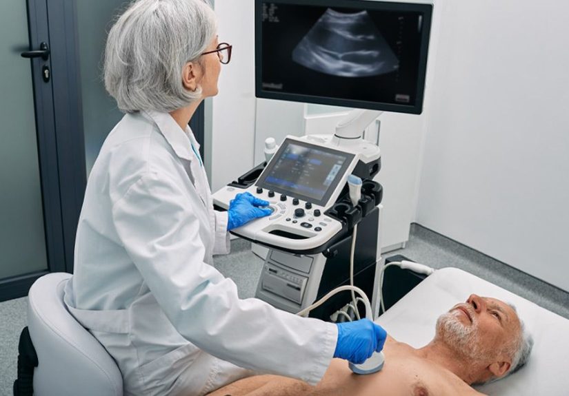
What Is an Echo?
An echo, short for echocardiogram, is a heart ultrasound. A technician or clinician moves a handheld probe over the chest with gel, and sound waves create real-time images of the heart. If an ECG is the heart’s electrical transcript, an echo is the live documentary.
What an echocardiogram shows
An echo helps doctors evaluate:
- The size and shape of the heart chambers
- How well the heart muscle pumps blood
- Valve function, including narrowing or leakage
- Blood flow through the heart using Doppler imaging
- Fluid around the heart
- Structural abnormalities, including some congenital heart conditions
This is why an EKG vs echocardiogram comparison is not really about which test is “better.” It is about what question needs answering. If the question is, “Is the rhythm abnormal?” an ECG may be the better first move. If the question is, “Why is there a murmur?” or “How well is the left ventricle pumping?” an echo becomes far more useful.
Different types of echo tests
Not all echoes are identical. The most common type is a transthoracic echocardiogram, which is the standard ultrasound done on the chest. A transesophageal echocardiogram, often called a TEE, uses a probe passed into the esophagus to get clearer images when doctors need a closer look. A stress echo evaluates how the heart performs during exercise or medication-induced stress. Doppler echo focuses on blood flow and pressure patterns.
In other words, “echo” is a whole family of tests, not just one.
ECG or EKG vs. Echo: The Biggest Differences
1. Electrical signals vs. moving images
The biggest distinction is simple. An ECG records electricity. An echo uses sound waves to make pictures. One listens to the heart’s wiring. The other watches the heart’s mechanics.
2. Speed vs. detail
An ECG is usually faster. It can be done in just a few minutes and is widely used in offices, urgent care centers, hospitals, and ambulances. An echo usually takes longer because it gathers much more structural and functional detail.
3. Rhythm problems vs. structural problems
ECGs are especially strong for rhythm and conduction issues. Echoes are especially strong for valve disease, chamber size, pumping strength, and anatomy. That is why doctors often use them together instead of choosing just one.
4. Snapshot vs. motion study
A routine EKG is brief, almost like a photo taken at one moment. An echo lets the clinician see the heart move in real time. That can matter a lot when trying to assess wall motion, valve opening and closing, or the direction of blood flow.
When Doctors Usually Choose an ECG First
In many real-world situations, an ECG is the first test because it is quick, inexpensive, and immediately useful. Common reasons include:
- Chest pain in the emergency room
- Palpitations or a racing heartbeat
- Fainting or near-fainting
- Suspected arrhythmia
- Preoperative heart assessment in some patients
- Checking pacemaker function
For example, if someone comes in with sudden chest pressure and sweating, the care team may order an ECG right away to look for signs of a heart attack or dangerous rhythm problem. Time matters here, and the EKG is built for speed.
When an Echo Becomes the Star of the Show
An echocardiogram often takes center stage when the concern is structural or functional rather than purely electrical. Common reasons include:
- A heart murmur that may suggest valve disease
- Possible heart failure
- Shortness of breath with suspected pumping weakness
- Known or suspected valve stenosis or regurgitation
- Evaluation after certain heart attacks
- Congenital heart disease assessment
Say a patient has swelling in the legs, fatigue, and shortness of breath. An ECG may still be ordered, but an echo can show whether the heart muscle is weak, whether the chambers are enlarged, or whether the valves are part of the problem. That is a different level of insight.
Why Doctors Often Order Both
One of the most useful things to understand about ECG or EKG vs. echo is that these tests answer different clinical questions. A cardiologist may want to know:
- Is the rhythm normal?
- Is there evidence of an old or active heart injury?
- How well is the heart pumping?
- Are the valves opening and closing properly?
- Is blood flowing in the right direction?
No single test covers all of that perfectly. An ECG and an echo together give a broader picture. One test may suggest a problem, and the other may clarify what that problem actually looks like.
This is especially true in conditions like heart failure, valve disease, atrial fibrillation, cardiomyopathy, and unexplained shortness of breath. The ECG may provide clues. The echo may show the consequences. Together, they tell a more complete story.
Which Test Is More Accurate?
That question sounds sensible, but it is slightly sneaky. Accuracy depends on what you are trying to detect. An ECG is more appropriate for rhythm abnormalities and certain signs of heart injury. An echo is more appropriate for evaluating heart structure, valve function, and pumping performance.
So asking whether an ECG is better than an echo is a bit like asking whether a thermometer is better than an X-ray. Better for what? If the problem is fever, use the thermometer. If the problem is a broken bone, please step away from the thermometer and call radiology.
Are These Tests Safe?
For most people, yes. A standard ECG is very safe and painless, though removing the adhesive pads can be mildly annoying if you have chest hair and strong opinions. A routine transthoracic echo is also considered safe and noninvasive, with no radiation involved.
More specialized versions of echo can carry additional considerations. A TEE may involve throat discomfort, sedation, and a small risk of complications. A stress echo may trigger symptoms during exertion or medication-based stress. That said, these tests are widely used and carefully supervised when clinically appropriate.
What Patients Usually Want to Know
Does an ECG hurt?
No. It is quick, painless, and usually over before your phone finishes checking for notifications.
Does an echo hurt?
A standard chest echo typically does not hurt, though the probe may press firmly in certain spots to get better images.
Can one test replace the other?
Usually not. They do different jobs. Sometimes one test is enough, but often both are used because they complement each other.
Which one checks for heart failure?
An ECG may suggest clues, but an echo is usually much more informative because it can show how well the heart is pumping and whether the chambers or valves are abnormal.
Which one is used for a murmur?
An echo is often the key test for a murmur because it can directly assess the heart valves and blood flow.
The Patient Experience: What It Actually Feels Like in Real Life
Here is where the conversation gets practical. Most people do not sit around thinking, “I hope my diagnostic workflow today is beautifully stratified.” They want to know what these tests feel like, how long they take, and whether they should be nervous.
The experience of getting an ECG is usually surprisingly ordinary. You check in, answer a few questions, and someone places electrode stickers on your skin. Then you lie still for a brief recording. That is it. The strangest part for many patients is how quick it is. The buildup feels dramatic because the word “heart” tends to get everyone’s attention, but the actual test is often over in a few minutes. Many people walk away thinking, “Wait, that was the whole thing?”
An echo feels more like a standard imaging visit. You are usually asked to lie on your side while a sonographer moves the transducer across your chest with gel. The room may be dimmed so the screen is easier to see. The test takes longer than an ECG, and the technician may pause in certain positions to capture better images. Sometimes there is firm pressure, especially between the ribs, but routine echoes are generally well tolerated. The most common post-test complaint is not pain. It is cold gel and a vague desire to know what the screen means immediately.
Emotionally, these tests can feel very different depending on why they were ordered. Someone getting an ECG as part of a routine check may feel mildly inconvenienced and mostly curious. Someone getting one in the emergency room may associate it with fear, urgency, and a flood of medical terms. The same goes for echoes. For one patient, it is a follow-up appointment. For another, it is the test that helps explain weeks of breathlessness or a newly discovered murmur.
Doctors also use the tests differently in ways patients can feel. An ECG often answers a fast question: Is there an immediate rhythm or ischemic concern? An echo often answers a deeper one: What is happening with the heart’s structure and function over time? That difference shapes the whole experience. The ECG can feel like a rapid checkpoint. The echo can feel like a more detailed investigation.
From a patient’s perspective, one of the most reassuring things is understanding that being sent for both tests does not automatically mean something terrible is going on. It often means the care team is being thorough. A person with palpitations may need the electrical snapshot of an ECG and the structural overview of an echo. A person with fatigue and shortness of breath may need both because symptoms overlap across many heart conditions.
The most relatable truth of all may be this: patients usually remember the waiting, the uncertainty, and the relief of finally getting answers more than the tests themselves. The ECG is quick. The echo is more visual. Neither is usually the hardest part. The hard part is the not knowing. Once you understand what each test does, the whole process feels less mysterious and a lot less scary.
Final Verdict: ECG or EKG vs. Echo
So, ECG or EKG vs. echo: what are the differences? The simplest answer is this: an ECG records the heart’s electrical activity, while an echo uses ultrasound to show how the heart looks and works. One is excellent for heart rhythm and electrical timing. The other is excellent for valves, chamber size, pumping function, and blood flow.
Neither test is “the better one” in every situation. They solve different pieces of the puzzle. If your doctor orders one, it is because they want a specific type of information. If your doctor orders both, it usually means they want the fullest picture possible. And when it comes to the heart, more clarity is rarely a bad thing.
Medical note: This article is for informational purposes only and should not replace professional medical advice, diagnosis, or treatment.