Table of Contents >> Show >> Hide
- What a PET scan actually measures (and why that matters in the liver)
- Why PET isn’t usually the first test for diagnosing primary liver cancer
- So… when is PET used in liver cancer?
- 1) Looking for cancer beyond the liver (staging and restaging)
- 2) Detecting recurrence after treatment (the “is it scar or is it cancer?” problem)
- 3) Evaluating treatment responseespecially in advanced disease
- 4) Helping with liver transplant decision-making in selected cases
- 5) Bile duct cancers (cholangiocarcinoma) and other hepatobiliary tumors
- 6) Clarifying indeterminate findings and guiding next steps
- PET/CT vs. PET/MRI: which one shows up in liver cancer care?
- Understanding “SUV” without needing a physics degree
- What to expect before and during the scan
- Limitations and “gotchas” in liver cancer PET imaging
- Where PET fits in a modern liver cancer workup
- Questions to ask your doctor (because you deserve a clear plan)
- Conclusion
- Experiences related to PET scans in liver cancer (patient & care-team perspectives)
If medical imaging were a group chat, CT and MRI would be the friends who always send crisp photos (“Here’s the tumor, here’s the blood vessel, here’s the problem”).
A PET scan is the friend who sends a vibe check: “Okay, but is that spot actually doing anything suspicious?”
In liver cancer, that “vibe check” can be incredibly helpfuljust not always in the way people assume. PET can be great for finding cancer that has traveled,
checking whether treatment worked, and clarifying confusing findings. But for spotting many primary liver tumors in the first place? PET is often more “sometimes” than “sure thing.”
Let’s break down how PET fits into the liver cancer toolbox, when it earns a starring role, and when it’s better as a supporting actor.
What a PET scan actually measures (and why that matters in the liver)
PET stands for positron emission tomography. Most PET scans used for cancer rely on a tracer called FDG (fluorodeoxyglucose),
which is essentially a tiny, radioactive “tag” attached to a glucose-like molecule. Because many cancer cells burn sugar faster than normal cells, FDG tends to collect
in areas with higher metabolic activity, which can light up on the scan.
PET is commonly paired with CT (PET/CT) and sometimes MRI (PET/MRI). The PET portion shows metabolic activity; the CT or MRI portion helps pinpoint the exact anatomy.
Think: “Where is it?” plus “What is it doing?”
Why PET isn’t usually the first test for diagnosing primary liver cancer
Here’s the twist: many primary liver cancersespecially hepatocellular carcinoma (HCC), the most common typedon’t consistently “drink” FDG the way
other cancers do. Some HCC tumors are very FDG-avid, while others are not. In practical terms, that means a PET scan can miss certain HCCs,
particularly well-differentiated tumors or very small lesions.
That’s why the heavy lifting for initial diagnosis and local tumor mapping is usually done by contrast-enhanced multiphase CT and
contrast-enhanced MRI. These tests can capture hallmark blood-flow patterns of liver tumors and help doctors decide whether a lesion looks like HCC
without always needing a biopsy. PET is typically not the first-line “find the primary tumor in the liver” test.
So… when is PET used in liver cancer?
PET’s value in liver cancer often shows up when the question shifts from “Is there a liver tumor?” to:
“Has it spread?”, “Is it back?”, or “Did treatment actually work?”
1) Looking for cancer beyond the liver (staging and restaging)
One of PET’s most common jobs in oncology is helping with stagingfiguring out how far cancer has spread. With liver cancer, staging matters a lot
because it can change the treatment plan entirely (surgery vs. transplant vs. localized therapy vs. systemic therapy).
PET/CT can be especially useful when doctors suspect extrahepatic disease (cancer outside the liver), such as lymph node involvement,
lung metastases, bone lesions, or other distant spread that might not be obvious on standard imaging.
2) Detecting recurrence after treatment (the “is it scar or is it cancer?” problem)
After treatments like ablation, embolization (including TACE), or radiation, the liver can look like it went through a tiny warbecause it did.
CT and MRI are still the standard tools for follow-up, but interpretation can be tricky when there’s inflammation, post-treatment change, or complex anatomy.
PET can sometimes help by highlighting areas of ongoing metabolic activity that look more like viable tumor than inactive scar tissue.
It’s not a magic truth serum (inflammation can also light up), but it can provide an extra layer of evidence when the picture is fuzzy.
Example: A patient has a treated lesion that’s stable in size but looks “weird” at the edges on MRI. If PET shows a focal hotspot in that same edge,
the team may lean toward additional treatment, a targeted biopsy, or closer surveillance.
3) Evaluating treatment responseespecially in advanced disease
PET can be useful for assessing response when the key question is “Are the cancer cells slowing down?” rather than “Is the tumor smaller?”
This can matter with systemic therapy, clinical trials, or complex cases where size alone doesn’t tell the whole story.
PET-based metrics (like changes in uptake) may provide prognostic clues in certain settings, though practices vary by institution and by the specific clinical scenario.
In everyday care, response assessment still relies heavily on CT/MRI, labs, and the patient’s overall clinical picture.
4) Helping with liver transplant decision-making in selected cases
For some patients with HCC, liver transplant can be curative. But transplant teams need to minimize the risk of transplanting someone whose cancer is
likely to recur aggressively. Because FDG uptake tends to be higher in more biologically aggressive tumors, PET positivity (when present) can contribute information
about tumor behavior and the likelihood of extrahepatic disease.
Important nuance: PET is not universally used for this purpose everywhere, and it is rarely the sole deciding factor. It’s one piece of a bigger puzzle
that includes tumor size/number, vascular invasion risk, AFP levels, response to bridging therapy, and overall health.
5) Bile duct cancers (cholangiocarcinoma) and other hepatobiliary tumors
“Liver cancer” isn’t one single disease. Cholangiocarcinoma (bile duct cancer) often behaves differently from HCC and may be more consistently FDG-avid.
In these cases, PET/CT can be used to help detect disease and evaluate spread, especially when planning treatment.
6) Clarifying indeterminate findings and guiding next steps
Sometimes imaging finds a lesion that’s suspicious but not definitive. PET may be used selectively to:
- Identify additional sites of disease that suggest metastasis
- Pick the best target for biopsy (the most metabolically active area can be the best “representative” sample)
- Support a decision to treat, observe, or pursue more imaging
PET/CT vs. PET/MRI: which one shows up in liver cancer care?
PET/CT is the workhorse. It’s widely available, fast, and excellent for whole-body surveys.
PET/MRI is less common, often more expensive, and typically available at specialized centersyet it can be attractive for liver imaging
because MRI provides superb soft-tissue contrast and specialized liver sequences.
In select casesespecially when metastatic staging is complex or when radiation exposure is a concernPET/MRI may be considered. But in most real-world workflows,
PET/CT is the default.
Understanding “SUV” without needing a physics degree
PET reports often mention SUV (standardized uptake value). SUV is a semi-quantitative estimate of how much tracer a spot absorbed compared with expectations.
Higher SUVs can suggest higher metabolic activity, which may be consistent with cancerbut SUV is not a diagnosis by itself.
SUV can be influenced by timing after injection, blood sugar levels, body composition, scanner calibration, inflammation, infection, and even how much you moved because
the waiting room chair was aggressively uncomfortable.
What to expect before and during the scan
Preparation
- Fasting: You’ll usually be asked not to eat for several hours beforehand. Water is typically allowed and encouraged.
- Avoid sugar/calories: Sugary drinks and snacks can interfere with FDG distribution and reduce scan quality.
- Diabetes planning: If you have diabetes, you may get special instructions about timing medications and meals.
- Tell your team about: pregnancy, breastfeeding, allergies (especially if contrast CT is planned), kidney issues, and all medications.
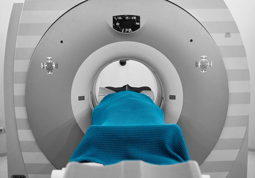
During the appointment
- You get an IV injection of tracer (often FDG).
- You wait while the tracer distributescommonly around an hour.
- You lie on the scanning table while the machine captures images.
The scan itself is painless. The most challenging part for many people is staying still and not thinking about how the ceiling tiles are suddenly the most fascinating
architectural feature ever invented.
Limitations and “gotchas” in liver cancer PET imaging
PET is powerful, but it has quirksespecially in the liver. Common issues include:
- False negatives: Some HCC tumors don’t show strong FDG uptake, so PET can miss themparticularly well-differentiated lesions.
- False positives: Infection, inflammation, healing tissue, and certain benign conditions can also show increased uptake.
- Small lesions: Very small tumors may not be detected due to resolution limits and background liver activity.
- Timing matters: Post-treatment inflammation can mimic disease if imaging is done too soon after therapy.
This is why PET results are interpreted in context, usually by nuclear medicine physicians and radiologists, alongside CT/MRI findings, labs (like AFP),
symptoms, and the overall clinical story.
Where PET fits in a modern liver cancer workup
A simplified way to think about it:
- CT/MRI: best for diagnosing and mapping tumors within the liver and guiding local treatment planning.
- PET (often PET/CT): best for evaluating whole-body spread, clarifying recurrence, and adding biologic context in selected cases.
In fact, many professional imaging pathways emphasize CT/MRI for routine primary liver cancer surveillance and follow-up, with FDG PET/CT used more selectively.
Your care team’s decision depends on tumor type, stage, prior treatments, and what question they’re trying to answer.
Questions to ask your doctor (because you deserve a clear plan)
- What specific question are we trying to answer with PET?
- Is PET/CT or PET/MRI recommended in my situationand why?
- How might the results change my treatment plan?
- What should I do about fasting and medications (especially diabetes meds)?
- If the scan is negative, what other tests will we rely on?
Conclusion
In liver cancer care, PET scans are less like a universal “find the tumor” button and more like a smart add-on when the situation calls for it.
They’re often used to look for disease outside the liver, investigate recurrence, and sometimes assess how active a tumor isespecially when standard imaging can’t fully
answer the question.
If your doctor recommends a PET scan, it usually means the team is trying to see the bigger picture: not just what a lesion looks like,
but what it’s doing and whether anything else is happening elsewhere. And in cancer care, that bigger picture can be the difference between a good plan and a great one.
Experiences related to PET scans in liver cancer (patient & care-team perspectives)
People often approach a PET scan with a mix of hope (“Please be clear!”) and dread (“What if it lights up everywhere?”). That emotional cocktail is normal.
When liver cancer is involved, the scan can feel even more high-stakes because treatment decisionslike whether a surgery is possible, whether transplant remains an option,
or whether therapy needs to changemay depend on what the PET/CT shows outside the liver.
One common experience is surprise at how much the preparation matters. Patients routinely report that the hardest part isn’t the scannerit’s the fasting,
the timing, and the logistics. If you’re juggling work, family, or diabetes management, being told “no food, no sugar, drink water, arrive early” can feel like a small
project plan disguised as a medical appointment. Many imaging centers provide detailed instructions, and when patients follow them closely, the scan quality is better,
which can reduce the chance of “we need to repeat this.” Nobody wants a sequel to an imaging appointment.
During the waiting period after the tracer injection, people often describe a strange calm: you’re just sitting there while the tracer circulates, trying not to move too much,
sometimes in a quiet room. Some patients bring music, an audiobook, or a meditation app. Others stare at a wall and become deeply philosophical about snack foods.
From the care-team side, that quiet hour matters because muscle activity can increase uptake in ways that complicate interpretation. The boring part is doing important work.
When results come back, patients can be thrown by the language. Words like “uptake,” “avid,” “hypermetabolic,” and “SUV” sound dramatic.
Clinicians often spend time translating: increased uptake can mean cancer, but it can also mean inflammation or healingespecially if the person recently had a procedure,
infection, or treatment. That’s why many care teams review PET results alongside CT/MRI and labs, and why tumor boards exist: a single scan rarely tells the whole story.
Patients who feel most confident afterward often say it wasn’t the scan that reassured themit was the explanation.
Another common experience is learning that a “negative” PET scan doesn’t always mean “no cancer,” particularly for certain hepatocellular carcinoma tumors that may not
light up strongly. Some patients feel confused“If I have liver cancer, why didn’t it show?”and that’s a fair question. Clinicians typically explain that different tumors
behave differently and that CT/MRI patterns in the liver are often more reliable for detecting certain primary lesions. In those cases, PET’s main value may be confirming
whether there’s disease elsewhere, not proving what’s in the liver.
On the flip side, when PET does show clear extrahepatic spread, patients describe a different kind of difficult clarity: the plan changes.
Sometimes that shift avoids an unnecessary surgery; sometimes it redirects treatment toward systemic therapy or clinical trials.
While that news can be hard, many patients later describe relief in knowing the care team isn’t treating only what’s visible in one organ while missing the bigger picture.
The “whole-body map” aspect of PET/CT can be emotionally heavy, but clinically valuable.
Finally, there’s a practical experience most people don’t expect: PET scans can be tiring. Not because they hurt, but because the appointment can take hours,
involves waiting, and can stir up anxiety. Many patients plan a low-key day afterward. Care teams often encourage hydration (as permitted) and normal routines once cleared,
because the tracer decays quickly and is eliminated over time. In the end, the scan is a moment in the larger storyone that can help guide smarter decisions,
even if it doesn’t answer every question all by itself.