Table of Contents >> Show >> Hide
- What Is an Eye and Orbit Ultrasound?
- Why Doctors Use Eye and Orbit Ultrasound
- Symptoms That May Lead to an Eye Ultrasound
- How to Prepare for an Eye and Orbit Ultrasound
- What Happens During the Procedure?
- Does an Eye Ultrasound Hurt?
- What Can Eye and Orbit Ultrasound Show?
- Benefits of Eye and Orbit Ultrasound
- Risks and Safety Considerations
- What Happens After the Test?
- Understanding Your Results
- Eye Ultrasound vs. Other Eye Imaging Tests
- When to Call a Doctor Urgently
- Practical Experiences and Patient Tips
- Conclusion
An eye and orbit ultrasound may sound like something that belongs in a sci-fi clinic, but it is actually a practical, painless imaging test used every day in eye care. Instead of radiation, it uses high-frequency sound waves to create images of the eyeball, the tissues behind the eye, and the bony socket area known as the orbit. In plain English: it helps doctors see what is happening when the eye refuses to give up its secrets.
This test is especially useful when a regular eye exam cannot show the back of the eye clearly. For example, dense cataracts, bleeding inside the eye, swelling, trauma, or cloudy eye structures can block a doctor’s view. An ultrasound can look beyond that cloudy “curtain” and help detect problems such as retinal detachment, vitreous hemorrhage, tumors, foreign bodies, inflammation, and orbital lesions.
Although the idea of having an ultrasound near your eye may make you blink just thinking about it, the test is usually quick, safe, and well tolerated. Most people finish it in minutes and return to normal activities right away. This guide explains the purpose of an eye and orbit ultrasound, how the procedure works, what conditions it may help diagnose, how to prepare, what the results may mean, and what risks to know before the scan.
What Is an Eye and Orbit Ultrasound?
An eye and orbit ultrasound, also called ocular ultrasonography, ophthalmic ultrasound, eye echography, or orbital ultrasound, is a diagnostic imaging test that uses sound waves to examine the eye and surrounding structures. A small handheld probe, called a transducer, sends sound waves into the eye area. These waves bounce back from tissues, fluids, and other structures, allowing a machine to create images or measurements.
The “eye” part of the test focuses on the eyeball itself, including the retina, lens, vitreous gel, optic nerve area, and internal eye structures. The “orbit” part looks at the tissues around and behind the eye, including muscles, fat, blood vessels, and the socket area. Together, they give doctors a clearer map of a very small but very important piece of real estate.
There are several types of ophthalmic ultrasound, but the two most common are A-scan and B-scan. They sound like grades on a school report, but thankfully no one is judging your eyeball’s homework.
A-Scan Ultrasound
An A-scan ultrasound is mainly used for measurements. It can measure the length of the eye, which is important before cataract surgery when doctors calculate the power of an artificial intraocular lens. It may also help measure certain tumors or other internal structures. The results appear as spikes or waveforms rather than a picture that looks familiar to patients.
B-Scan Ultrasound
A B-scan ultrasound creates a two-dimensional image of the eye and orbit. This is the type many people think of when they imagine an ultrasound picture. It is often used when the doctor needs to evaluate the back of the eye, especially if something blocks the view during a standard exam.
Ultrasound Biomicroscopy
Ultrasound biomicroscopy, often called UBM, uses higher-frequency ultrasound to examine the front portion of the eye in fine detail. It may be used to assess narrow angles, angle-closure glaucoma risk, ciliary body problems, certain cysts, trauma, or lens-related concerns. Because it gives detailed images of the anterior segment, it can be helpful when other imaging tests do not answer the question.
Why Doctors Use Eye and Orbit Ultrasound
The main purpose of an eye and orbit ultrasound is to help diagnose or evaluate eye conditions when a direct view is limited or when measurements are needed. Your eye doctor may recommend the test after symptoms, injury, an abnormal exam, or before certain procedures.
One of the most common reasons for the test is a cloudy view into the eye. In a healthy exam, an ophthalmologist can usually look through the pupil and examine the retina. But when cataracts, bleeding, severe inflammation, corneal clouding, or other problems block the view, ultrasound becomes the eye-care equivalent of turning on headlights in fog.
Common Reasons for an Eye Ultrasound
An eye and orbit ultrasound may be used to check for:
- Retinal detachment: A serious condition in which the retina pulls away from the back wall of the eye.
- Vitreous hemorrhage: Bleeding into the gel-like substance inside the eye.
- Posterior vitreous detachment: Separation of the vitreous gel from the retina, which can cause floaters or flashes.
- Eye tumors: Including intraocular melanoma or other masses.
- Foreign bodies: Small objects that may have entered the eye after trauma.
- Orbital lesions: Abnormal growths, swelling, or tissue changes behind the eye.
- Inflammation or infection: Especially when swelling affects deeper structures.
- Optic nerve concerns: In some cases, ultrasound can help assess the optic nerve sheath.
- Cataract surgery planning: A-scan measurements may help determine lens implant power.
- Eye trauma: When used carefully and only when globe rupture is not suspected.
For patients, the most important point is that ultrasound is not usually ordered “just because.” It is ordered when it can answer a specific question that affects diagnosis, treatment, or surgical planning.
Symptoms That May Lead to an Eye Ultrasound
Your doctor may recommend an ocular ultrasound if you have sudden or unexplained eye symptoms. These symptoms do not always mean something dangerous is happening, but they should be evaluated promptly because some eye problems are time-sensitive.
Symptoms that may lead to the test include:
- Sudden vision loss or a curtain-like shadow over vision
- New flashes of light
- A sudden increase in floaters
- Eye pain after injury
- Blurred vision that cannot be explained by glasses
- Swelling around the eye
- Bulging of the eye
- Concern for bleeding inside the eye
- A mass or abnormal finding during an eye exam
- Poor view of the retina because of cataract or cloudy media
Some of these symptoms, especially flashes, floaters, sudden vision loss, or a dark curtain in the visual field, can be associated with retinal detachment. That is not a “wait and see” situation. It is more of a “call the eye doctor now” situation.
How to Prepare for an Eye and Orbit Ultrasound
Preparation is usually simple. In many cases, no fasting, sedation, or special medication is needed. You can eat normally, take your regular medicines unless your doctor says otherwise, and wear comfortable clothing. The eye itself gets all the attention; your outfit does not need to impress the ultrasound machine.
Before the Test
Your care team may ask about your symptoms, eye history, surgeries, injuries, allergies, and current medications. If the test involves direct contact with the eye, numbing drops may be used. If the probe is placed over the closed eyelid, gel is applied to help transmit sound waves.
If you wear contact lenses, ask whether you should remove them before the exam. This is especially important if numbing drops are used. You may be told not to put contacts back in until the drops wear off and your eye feels normal again.
Tell Your Doctor About Eye Trauma
This step is important: tell the doctor if you recently had an eye injury, especially a sharp injury, high-speed impact, chemical injury, or severe blunt trauma. If there is concern for an open-globe injury or globe rupture, pressure on the eye can be dangerous. In that situation, doctors may choose other imaging, such as CT, and involve ophthalmology urgently.
What Happens During the Procedure?
The procedure depends on the type of ultrasound being performed. Most eye ultrasounds are done in an ophthalmology office, imaging center, hospital, or emergency department. The test is usually short, often taking only several minutes once everything is set up.
Step 1: You Are Positioned Comfortably
You may sit in a chair or lie back on an exam table. The technician, ophthalmologist, or trained clinician explains what will happen. You may be asked to close your eyes, look in certain directions, or stay still while the probe collects images.
Step 2: Gel or Numbing Drops Are Applied
For a scan over the eyelid, a clear ultrasound gel is placed on the closed eyelid. The gel may feel cool and a little strange, but it is harmless and wipes off afterward. For some direct-contact scans, numbing eye drops may be used so the surface of the eye does not feel irritated.
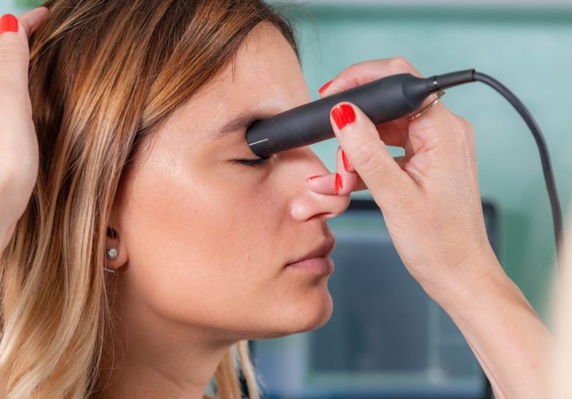
Step 3: The Probe Is Moved Gently
The probe is moved gently over the closed eyelid or near the eye surface. The clinician uses light pressure and adjusts the angle to capture images from different directions. You may be asked to look up, down, left, or right. This helps the examiner see how internal structures move, which can be useful when distinguishing between retinal detachment, vitreous detachment, and other findings.
Step 4: Images or Measurements Are Recorded
The ultrasound machine records images, measurements, or waveforms. In a B-scan, the clinician evaluates the shape and movement of structures inside the eye. In an A-scan, measurements may be taken for biometry or tumor assessment.
Step 5: The Gel Is Cleaned Off
After the scan, the gel is wiped away. If numbing drops were used, your eye may feel temporarily different until sensation returns. Avoid rubbing the eye while it is numb, because your eye’s “ouch alarm” may be temporarily offline.
Does an Eye Ultrasound Hurt?
Most people describe an eye and orbit ultrasound as painless. You may feel cool gel, mild pressure, or brief awkwardness because someone is working very close to your eye. But the test should not be painful. If you feel pain, pressure, dizziness, nausea, or significant discomfort, tell the clinician immediately.
People who are sensitive about anything near their eyes may feel nervous. That is normal. The good news is that the test is quick, and the person performing it is trained to use gentle technique. You can ask them to explain each step before they do it. Sometimes knowing what is coming helps the nervous system stop acting like a raccoon trapped in a garage.
What Can Eye and Orbit Ultrasound Show?
An ultrasound can show a wide range of abnormalities, depending on the reason for the test. It may help identify whether the retina is attached, whether blood is present inside the eye, whether a mass exists, or whether an orbital structure looks abnormal.
Retinal Detachment
Retinal detachment occurs when the retina separates from the back of the eye. On ultrasound, it may appear as a membrane-like structure attached near the optic nerve. Because retinal detachment can threaten vision, fast diagnosis and treatment are important.
Vitreous Hemorrhage
Vitreous hemorrhage means blood has leaked into the clear gel inside the eye. This can happen from diabetic eye disease, trauma, retinal tears, vascular problems, or other causes. Ultrasound can help determine whether the retina is still attached when blood blocks the doctor’s view.
Eye Tumors or Masses
Ultrasound may help evaluate intraocular tumors, including melanoma, by showing size, shape, location, and internal characteristics. It does not replace all other testing, but it can provide valuable information for diagnosis and monitoring.
Foreign Bodies
After an injury, ultrasound may sometimes help detect foreign material in or near the eye. However, if an open-globe injury is suspected, doctors must avoid putting pressure on the eye and may choose other imaging first.
Orbital Problems
Orbital ultrasound may help evaluate swelling, inflammation, cysts, tumors, muscle enlargement, or other tissue changes around the eye. It can also be useful when symptoms involve bulging, pain with eye movement, or unexplained swelling.
Benefits of Eye and Orbit Ultrasound
Eye ultrasound has several advantages. It is fast, does not use ionizing radiation, and can provide useful information when other parts of the eye exam are limited. It can also be performed at the bedside in certain emergency settings, which is helpful when time matters.
Key benefits include:
- No radiation: Ultrasound uses sound waves, not X-rays.
- Quick results: Images are often available immediately for the clinician to interpret.
- Useful through cloudy structures: It can evaluate the back of the eye when cataracts or bleeding block the view.
- Dynamic imaging: The examiner can watch how structures move when the eye moves.
- Helpful for measurements: A-scan ultrasound can measure eye length and support cataract surgery planning.
- Widely available: Many ophthalmology practices and hospitals have access to ocular ultrasound.
Risks and Safety Considerations
For most people, an eye and orbit ultrasound is considered very safe. It is noninvasive or minimally contact-based, does not involve radiation, and rarely causes side effects. Still, no medical test should be treated like a casual selfie. Technique matters, training matters, and the reason for the test matters.
Possible Minor Side Effects
Minor effects may include temporary irritation, watery eyes, mild redness, or a strange sensation if numbing drops are used. These usually resolve quickly. Some people may feel anxious during the test because the probe is close to the eye.
Pressure on the Eye
The main safety concern is pressure. The probe should be used gently, especially if the eye has been injured. Too much pressure can cause discomfort and may be risky in certain trauma situations. Skilled clinicians use plenty of gel, light contact, and careful positioning.
Suspected Globe Rupture
If doctors suspect a globe rupture, meaning the outer wall of the eyeball may be open or torn, ultrasound is usually avoided or used only with extreme caution by specialists. Pressure on a ruptured eye can worsen injury. Warning signs may include severe pain after trauma, distorted eye shape, leaking fluid, decreased vision, or a history of sharp or high-speed injury.
Rare Reflex Reactions
In rare cases, pressure on the eye can trigger a reflex that causes nausea, lightheadedness, or a slower heart rate. This is uncommon, but it is one reason patients should speak up if they feel unwell during the exam.
What Happens After the Test?
After an eye and orbit ultrasound, most people can return to regular activities right away. If gel was used, it will be wiped off. If numbing drops were placed in the eye, you may be advised not to rub your eye until normal feeling returns. If you wear contact lenses, ask when it is safe to put them back in.
Your doctor may discuss the findings immediately or schedule a follow-up visit. In urgent situations, such as suspected retinal detachment, you may be referred quickly to a retina specialist or ophthalmic surgeon. In less urgent cases, results may guide monitoring, medication, additional imaging, or surgical planning.
Understanding Your Results
Results may be described as normal or abnormal, but the meaning depends on your symptoms and eye history. A normal ultrasound may show that the retina appears attached, no mass is visible, and the internal structures look as expected. However, a normal scan does not always rule out every possible eye condition. Your doctor will interpret the ultrasound alongside your exam, symptoms, and other tests.
An abnormal result may show bleeding, detachment, swelling, inflammation, a foreign body, tumor, or orbital abnormality. Try not to panic if the doctor says something was “seen” on the scan. Ultrasound is a clue-finding tool, not an instant life sentence. Many findings require follow-up testing, specialist evaluation, or repeat imaging before a final diagnosis is made.
Eye Ultrasound vs. Other Eye Imaging Tests
Eye and orbit ultrasound is only one tool in the diagnostic toolbox. Other tests may include optical coherence tomography, fundus photography, fluorescein angiography, CT, or MRI. Each test has strengths and limitations.
Optical coherence tomography, or OCT, creates detailed images of the retina but requires a reasonably clear path through the eye. If cataracts or bleeding block the view, ultrasound may be more useful. CT scans are often used after trauma, especially when doctors are concerned about fractures, foreign bodies, or open-globe injury. MRI may be used for some orbital or neurologic concerns, but it is not used when certain metallic foreign bodies are possible.
In many cases, these tests do not compete with each other. They work as a team. Ultrasound may answer one question, OCT another, and CT another. The goal is not to collect medical images like trading cards; it is to choose the right test for the right problem.
When to Call a Doctor Urgently
Seek urgent eye care if you develop sudden vision loss, a curtain or shadow in your vision, new flashes, many new floaters, severe eye pain, eye injury, chemical exposure, or swelling with fever. These symptoms can signal serious eye conditions that need fast attention.
You should also call your doctor if symptoms worsen after an exam or if you have persistent redness, pain, or vision changes. Eye problems can move quickly, and early treatment often protects vision better than waiting for things to “calm down” on their own.
Practical Experiences and Patient Tips
For many patients, the biggest challenge of an eye and orbit ultrasound is not pain. It is imagination. The brain hears “ultrasound near your eye” and immediately begins writing a horror movie starring a cold probe and dramatic background music. In real life, the experience is usually much calmer. Most patients feel gel on the eyelid, gentle movement, and mild pressure. The test often ends before the nervous part of the brain has finished complaining.
A common experience is surprise at how simple the setup is. You may expect a large machine, bright lights, and a complicated process. Instead, the clinician may ask you to lean back, close your eye, and hold still while they apply gel. If the scan is done over the eyelid, you do not have to stare at the probe. That alone makes the test easier for people who dislike objects coming toward their eyes.
Another helpful tip is to relax your forehead and eyelids. People often squeeze their eyes shut when they are nervous. That is understandable, but it can make the exam harder. Think of gently resting your eyelids rather than clamping them like a vault door. Slow breathing helps. Try inhaling through your nose and exhaling slowly through your mouth while the clinician scans.
If you are having the test because of floaters, flashes, or sudden blurry vision, bring a clear description of what happened. For example, tell your doctor when symptoms started, whether they appeared suddenly or gradually, whether you saw a curtain-like shadow, and whether you had recent trauma. These details help the clinician match the ultrasound findings to the most likely cause.
If you are being evaluated before cataract surgery, your experience may be slightly different. The focus may be on measurements rather than searching for bleeding or detachment. You may hear terms such as axial length, lens calculation, or biometry. These measurements help the surgeon choose the right artificial lens implant. It is a little like tailoring a suit, except the suit goes inside your eye and nobody asks whether you prefer navy or charcoal.
Patients with anxiety should tell the care team before the scan begins. A good clinician can explain each step, pause when needed, and remind you that the test is brief. You are not being dramatic if eye procedures make you uneasy. Eyes are sensitive, and most people are naturally protective of them. Communication makes the appointment smoother.
After the test, ask what happens next. Useful questions include: “Did the ultrasound show the retina clearly?” “Is there any sign of retinal detachment?” “Do I need to see a retina specialist?” “Are my symptoms urgent?” “Should I avoid any activities today?” “When should I call if symptoms change?” These questions turn the ultrasound from a mysterious scan into a practical plan.
It is also wise to avoid driving yourself if your appointment may include dilating drops or if your vision is already significantly affected. The ultrasound itself usually does not impair vision, but the full eye exam around it might. Sunglasses can help if your pupils are dilated afterward and the outside world suddenly looks like it turned up the brightness setting.
Finally, remember that an eye and orbit ultrasound is not a treatment. It is a diagnostic tool. The value of the scan comes from what it helps your doctor decide next. Sometimes the result is reassuring. Sometimes it leads to urgent treatment. Sometimes it points toward additional testing. In every case, the test is designed to protect one of your most precious senses: sight.
Conclusion
An eye and orbit ultrasound is a safe, useful imaging test that helps doctors evaluate the eye and surrounding socket when a standard exam does not provide enough information. It can help detect retinal detachment, vitreous hemorrhage, tumors, foreign bodies, inflammation, and orbital problems. It also plays an important role in measuring the eye before certain surgeries, especially cataract surgery.
The procedure is usually quick and painless. Most people need little preparation and can return to normal activities afterward. The main safety concern is suspected globe rupture after trauma, because pressure on an injured eye can be dangerous. That is why it is important to tell your doctor about any recent injury before the scan.
If your doctor recommends an ocular ultrasound, it does not automatically mean something serious is wrong. It means they want a clearer view, better measurements, or more information before deciding what to do next. In eye care, clarity is everythingand sometimes sound waves are the best way to get it.